The Lizard Log

The Langkilde Lab in Action


Leave a comment

A Langkilde lab #reviewforscience

While looking up ideas for how to run some of my immune assays in the field, I stumbled across the twitter hashtag, #reviewforscience, where scientists leave reviews for common, everyday items they use in their research. So, in the interest of sharing how our lab has repurposed some everyday items for science, I thought I’d do a few reviews of my own.

Dental flossdental floss

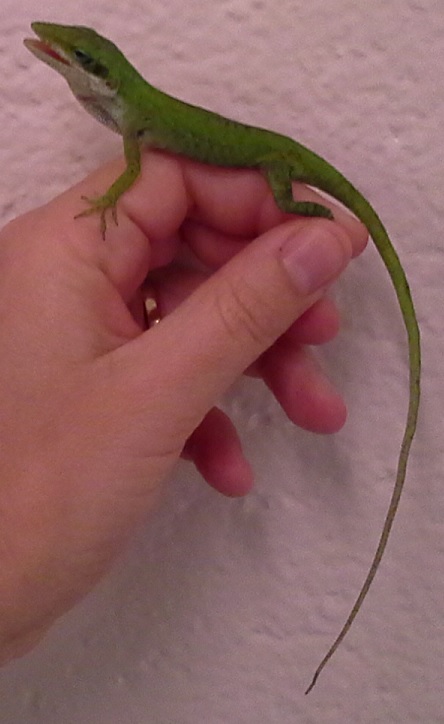
As has been noted by other esteemed colleagues, dental floss is excellent for making nooses when capturing lizards. Just tie the noose on the end of a cheap fishing pole to reach lizards high up in trees!


Plastic Sterilite tubs

IMG_20170802_114752001_HDRIMG_20170802_114803995

Work well for individual or group-housed adult fence lizards. Adding clean, damp sand substrate makes an ideal habitat for females ready to lay and bury their eggs. Smaller versions can be used for hatchlings and juvenile fence lizards. Easily sterilized in standard cage washes! Just remember to wrap in opaque paper before use to prevent adult males from seeing one another.


24 hour plug-in timerstimer

Work great for timing basking lights to match up with daylight hours outside, to maintain the circadian rhythms of captive-housed fence lizards. Make sure you get the type with 2 grounded outlets!


Plastic deli containersIMG_20180112_111835361

So many uses in a lizard lab! Can be used to hold lizards, crickets, and eggs for weighing. Great for transporting hatchlings from the lab to the field! When filled with damp vermiculite, they make an excellent place to incubate fence lizard eggs!


Thermocouple thermometer img_20170221_131145

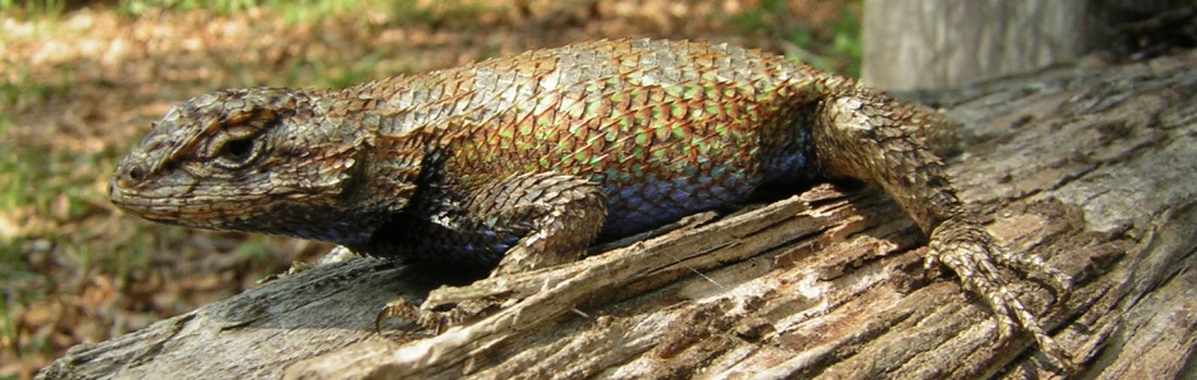
Like other scientists, we’ve found that a thermocoupler probe “fits neatly inside a lizard’s cloaca” for measuring body temperature.


Bug vacuumbug vacuum

Works great for collecting leftover crickets in order to track how many were eaten. Fairly quiet, and less stressful to fence lizards than reaching in to hand-capture the crickets, as well as being much less time consuming! And crickets are undamaged, so can be used again in later feedings (hand capture of crickets often results in squished crickets). Long-lasting battery, can vacuum over 100 cages before needing to be recharged.


Honey

honeyIMG_20180123_120223668

Surprisingly effective organic glue for attaching deceased fire ants to live crickets for a fence lizard feeding study. I wanted the fence lizards to consume fire ants without being stung, which meant the fire ants needed to be euthanized first. But fence lizards won’t consume unmoving prey, so I had to attach the fire ants to a living insect. Using <5µl of honey, I was able to stably attach 10 fire ants to each cricket, and the cricket was still able to move about freely.


Tea strainertea strainer

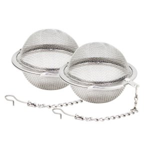
Great container to hold isoflurane-soaked cottonballs for lizard anesthesia. The wire mesh allows the lizard to breath in the isoflurane, but holds in the cotton ball to avoid direct contact with the animal. Have not tried to use tea strainers with fire ants. Can also be used to strain particulate matter out of feces when performing fecal parasitology tests.


Plaster of Parisplaster of paris

Used in the production of fake lizard models for predation studies. Easy to use, and held up well to short-term exposure to the elements.


White duct tapeduct tape

Works well to make dry-erase labels for tubs, in order to quickly label and re-use for weighing crickets and lizards.


Sous vide cookersous vide cooker

I’m thinking of using one of these this field season as an inexpensive, travel-friendly alternative to a water bath. When in the field, just clip this onto the side of a container full of water, plug into a wall outlet or portable car charger, set the temperature you want the water at, and go! No need to transport an expensive, bulky water bath.


Leave a comment

Immunapalooza

Not all of the lab went south this summer; Kristen (one of our awesome lab undergrads) and I stayed at Penn State most of the summer, working on immune assays.  Kristen was the recipient of the Erickson Discovery Grant, and spent much of her summer on an independent research project, which involved measuring the effects of corticosterone (CORT) on the cell-mediated immunity (i.e. one way the body responds to a toxic or foreign substance) of eastern fence lizard females. She was also trying to determine if the lizards’ life history (whether they were from sites with or without fire ants) affected their immune function or interacted with the CORT treatment. Kristen just recently gave an excellent talk on her research at the Three Rivers Evolution Event (TREE) on Sept. 9th, where she was one of the only undergraduates to present a talk.

We also spent a lot of time this summer developing, improving, and validating several different immune assays for use in fence lizards, including ELISA assays for measuring anti-fire ant antibodies (IgY and IgM), complement function, natural antibodies, and the activity levels of heterophils (a type of immune cell that kills bacteria). Work on the assays for IgY, complement function, and natural antibodies is ongoing, but the IgM and heterophil activity assays are ready to be used.

The IgM ELISA assay was developed to work with as little as 10μl of plasma, and accurately detected anti-fire ant antibodies in a pool of plasma of lizards from Alabama, where the lizards are regularly exposed to fire ants. It did not detect any antibodies in a pool of plasma of lizards from Tennessee, at sites which have not yet been invaded by fire ants. The next step is to test the plasma of individual lizards from different sites, to see what proportion of lizards in various invaded sites have actually developed IgM antibodies to fire ants. Once the IgY assay is working, we should be able to better characterize the antibody response of the lizards to fire ants, and see if this helps them recover faster from fire ant stings.

IgM in the plasma of Alabama lizards

The higher the proportion of plasma from invaded (Alabama) lizards, the higher the signal from the IgM antibody.

Our heterophil activity assay is based off the assay described in Merchant, Williams, and Hardy (2009) for use in American alligators. To account for the much smaller blood volume of fence lizards, I altered the assay to work with 10μl of whole blood, and validated it in this species. This assay specifically tests for the presence of superoxide radicals, which are produced by heterophils as part of the oxidative burst used to kill bacteria and other organisms. When heterophils are more active (either because there are more heterophils or because the existing heterophils have been stimulated by something), the amount of superoxide in the blood increases. As part of the validation, we ran the assay with pools of blood treated with superoxide dismutase, which destroys superoxide, to test that the signal is actually caused by superoxide. We also ran blood with and without a stimulant of heterophil function, to determine if the signal reliably increases when heterophils are more active. The signal reliably decreases when inhibited by superoxide dismutase, and reliably increases when stimulant is added, indicating that this is a reliable test of heterophil function.

We also did a little bit of work optimizing the natural antibody test, increasing the sensitivity of the test so that it will work with less lizard plasma. And we also found a promising lead for testing alternative pathway complement function in fence lizards.

Aside from all the immunology work, we also got out into the field up here in Pennsylvania a little bit, although we didn’t find many lizards. All in all, it was a fun, productive summer.


Leave a comment

Lizard poop and the parasites who love it

A lot of my work in the lab involves assessing the health and well-being of our fence lizards under different conditions, including their parasite burdens. Parasite infestation can vary with immune status, stress, or external factors such as predation (for example, lizards in fire ant invaded areas have fewer ectoparasites). Ectoparasite (ticks, mites, etc.) load is easy to assess, as we count them on each lizard shortly after capture. Internal parasites are a bit trickier, but one method commonly used in veterinary medicine is to collect their feces, and check it for intestinal parasites and eggs.

There are several different methods of testing for intestinal parasites, including direct smears, qualitative fecal flotations, and quantitative fecal egg counts. Direct smears are the simplest method, involving looking at fecal smears directly under a microscope, but they are also the least sensitive, and often don’t show any results. The most sensitive method is qualitative fecal flotations, the method of choice if you want to see all the possible parasites an organism may have in their feces.
The basic idea behind a fecal flotation is a feces sample is mixed with a solution denser than the parasite eggs you are looking for. The mixture is then spun in a swinging-bucket centrifuge. Due to the parasite eggs having a lower density than the solution, they float to the top of the tube while being centrifuged, and collect on a cover slip on the top of the tube. This results in most of the parasite eggs in the fecal sample being concentrated onto the cover slip for easy viewing.
Unfortunately, the fecal flotation method, while a great way to learn how many different types of parasites are in a fecal sample, does not tell you how many individual eggs are in each gram of feces. Such comparisons are important in veterinary medicine in order to tell if a treatment is working, and is important to us in the lab for comparing fecal egg loads between experimental groups. This is where quantitative fecal egg counts become useful. While less sensitive than fecal flotations (they may not identify lower-level infestations of parasites), fecal egg count methods can tell us how many eggs are in each gram of feces. To do this, we precisely dilute a set amount of feces into flotation solution, and mix it thoroughly. The mixture is then placed in a special slide, called a McMaster, and read after 5 minutes, using the grid on the slide.
mcmaster-counting-slides-triple-chamber

The McMaster slide we use

I’ve had some challenges adapting these methods to use in our fence lizards, as both fecal flotation and fecal egg counts require more feces than a lizard normally produces, but I have gotten some interesting results, mostly a variety of strongyle and coccidia eggs.


Leave a comment

Much Anole About Immunology

While most of the lab has been down in Alabama, I’ve spent a good part of this summer back at Penn State, working with a species I’ve never used before – the green anole (Anolis carolinensis).

20160727_084137_edit

Isn’t he cute?

There is a test we’d like to use in our fence lizards, called the phytohemagglutinin (PHA) skin test. It involves injecting the pad of a rear foot with a small amount of PHA, which stimulates part of the immune system, and then measuring the swelling that occurs. This swelling is small, and temporary, abating in a few days with no lasting damage. But the level of swelling can provide information about the lizards’ immune function.

20160727_084019

A confused anole having its rear foot measured with calipers.

Unfortunately, while this test has been used in humans, birds, rats, and even amphibians, it has not yet been validated in any reptile species. Ideally I would validate the test in our species of interest, the eastern fence lizard, but I needed a larger number of lizards than we can reasonably catch. So, instead, we decided to purchase some green anoles for this project.

In addition to seeing if the PHA test works in reptiles, we’re also trying to determine if the type of PHA used makes a difference, as there are many different formulations of PHA used, and each formulation may have a different effect. I’m also determining exactly what the immune reaction to the different PHA formulations are, and how this evolves over time after the injection.


Leave a comment

Veterinary School:  Is it worth it?

As the lab’s resident veterinarian, I’m frequently asked questions about getting a Doctor of Veterinary Medicine degree. Things like, “What were you thinking?” and “Does thinking about your student loans keep you up at night?” I thought I would cover a few of these topics here in the lab blog.

To start with, to anyone who is considering applying to veterinary school:  It is NOT a smart economic decision. The cost of a Doctor of Veterinary Medicine (DVM or VMD) degree, on average, barely breaks even over the course of a man’s career, and actually slightly decreases the lifetime earnings of a woman. Given that 90% of veterinary students graduate with student loan debt (how did 10% of students manage to graduate without any debt??? So jealous.), and the average student loan burden is over $160,000, this is not surprising. Compounding the financial strain is the relatively low starting salaries of veterinarians, only around $67,000 a year if they start working full-time immediately, rather than pursuing further training such as a PhD, internship, or residency. And there doesn’t seem to be any advantage to attending a more expensive school; starting salary is in no way correlated to student loan debt or the cost of tuition.

Vet student debt to income

The direness of the financial situation of course varies considerably by field, with food animal and equine veterinarians having the worst debt to income ratio, and vets working in industry having the best. Veterinarians working in academia (like me!) fall somewhere in between, but we at least have a decent chance of paying back our student loans in a reasonably timely manner. Even if I do often feel like this:

Student loan debt

There is a pervasive myth that, in spite of the heavy debt load and relatively poor earning potential, there is a national shortage of veterinarians, so it is at least a good field to go into for job security. This, unfortunately, is not true. While the number of job openings for veterinarians are predicted to increase by 9% from 2014-2024, the number of graduating veterinarians has been rising by 1.8% per year for the last 30 years (we currently graduate ~3000 new DVMs per year), a trend which is expected to continue. And a 2012 report by the National Research Council (NRC) showed that there is no national shortage of veterinarians, except in some rural areas. This is also consistent with the findings of the 2013 AVMA veterinary workforce report, which showed that 12.5% of the veterinary capacity to provide services is going unused (this does not mean that 12.5% of veterinarians are unemployed or underemployed, just that as a whole, 12.5% of the potential services that could be provided by veterinarians around the country are going unused each year). However, this is not necessarily bad – there is also not an extreme surplus of veterinarians, and unemployment in the field is lower than the national average, only 3.4%. And having a moderate surplus of capacity means we can better handle emergencies such as disease outbreaks.

So, given that we’ve established veterinary school is a terrible, terrible, terrible financial decision, why do people still get the degree? And why am I among them? Well, for most people I think it’s more of a calling than a career. Most veterinarians desperately love their job, love caring for animals, and consider the incredible financial burden to be worth it if it means they get to spend every day saving lives. In my case, I never intended to be a full time practicing veterinarian. My plan was always to obtain both a DVM and a PhD, and work as a researcher. And, as I said earlier, the financial outlook for a veterinarian in academia is much better than for most practicing veterinarians; my salary is likely to be higher, and I can at least start paying off my student loans while working on my PhD.  The big payoff to the veterinary degree, for me, is a detailed knowledge and understanding of a wide variety of animals. I focused a lot of my study on interspecies comparisons, and how to extrapolate information from one species to be able to apply it in another. This has proven immensely useful in my academic career; even though I haven’t worked extensively with reptiles or amphibians prior to entering this lab, my knowledge in a wide variety of other species is still applicable. Medical training is also much more thorough, in general, than PhD classes are; there is no way I could have gained this depth of knowledge without having obtained a medical degree of some kind. And veterinarians are uniquely well-suited to answering questions about comparative anatomy and physiology, and to determining how likely a disease or medicine is to work in one species versus another (compare this to human medical doctors, who are taught the same things as veterinarians, but in only one species). This is invaluable in efforts to determine how wide-ranging an effect a research finding might have. For example, research done by Gail shows that stress causes immunological changes in the Eastern fence lizard (Sceloporus undulatus). My veterinary training makes me well-suited to developing hypotheses as to why this might be (questions based on what parts of the immune system are affected, what the time line of the changes are, etc.), and how likely this change is to be something that would occur in other species in the same situation (i.e., would a squirrel/snake/fish/etc. experience the same immune system changes when exposed to the same type of stress?). It also helps me explain some of our findings better, like how stress might cause changes in certain portions of the immune system, but cause no changes, or even opposite changes, in other parts.

While there has been a push to include human medical doctors in research using human subjects, there has not been as concerted an effort to do the same with animal subjects. All animals used in research are of course overseen by veterinarians, but usually their role is limited to the care and compassionate use of the animal subjects. I personally feel that most fields of animal research would benefit greatly from more veterinary input in developing hypotheses to test, and in study design and interpretation. One of my career goals is to illustrate how veterinary knowledge and training can benefit the research community, and encourage more scientists to obtain veterinary training, or encourage more veterinarians to participate in research.


Leave a comment

Return to Research Land!

Hi, I’m Caty Tylan, one of the new Ph.D. students in the Langkilde Lab. Since this is my first post, it will mostly be an introduction. Short version: I’m a huge science nerd who reads, cooks, and plays a LOT of board games in my spare time. I have 1 adorable husband, and 3 adorable pets – a cat, a rabbit, and a very large dog.

Me with our giant puppy.

He luuuuuurrrvves me.

He’s a little needy.

Our tiny Holland Lop.

My nieces and nephews LOVE our bunny.

The cat is not amused.

But she is adorable.

I just graduated with my D.V.M. from Purdue University, but as the title of this post implies, I spent a lot of time in various labs before going to veterinary school, and I am excited to be doing research again! I obtained my B.S. in Biology from Drexel University, where I worked in a couple of different ecology laboratories, and also had the opportunity to work at Merck & Co., Inc. for my cooperative education experiences. After undergraduate I moved to Indiana and worked as a laboratory technician in the Bacteriology department of the Purdue University Animal Disease Diagnostic Laboratory. During this time, I was lucky enough to work with a wide variety of species, which I loved. However, I wanted to know more about how they worked on a basic biological level before doing more research with them, so I decided to attend veterinary school. I was initially co-enrolled in a Ph.D. program, where I rotated through a couple more laboratories, but I eventually left the Ph.D. program due to conflicts with my D.V.M. program.

While completing my D.V.M. I started a project to test out a novel euthanasia method for use in domestic ducks. There is currently no good method for euthanizing large numbers of ducks, which is sometimes needed in cases such as disease outbreaks. The method that we’ve been testing out involves injecting alcohol into the duck’s brain, which has proven to be arguably the least traumatic method for the ducks.. The brain does not have the correct type of receptors to feel pain from the needle’s insertion, so the ducks experience no more pain than if they were having their blood taken, quickly followed by unconsciousness and death.  We are currently working on publishing our results.

I have minimal background in herpetology, having done some field work with turtles during undergraduate, but I’m excited to apply my medical knowledge to enhancing the study of fence lizards and other animal species in the Langkilde lab. I’m starting out focusing on the study of stress and separating out the effects of glucocorticoid hormones from the other mediators of stress in fence lizards.